What I Treat?
1. Knee Joint
1.1 Arthritis

Knee arthritis is a common condition that affects the cartilage in the knee joint. The cartilage is what protects your bones from rubbing against each other.
Knee arthritis usually starts with some wear and tear of the cartilage, which can lead to pain and stiffness in the joint. The condition can be caused by injury or overuse, and it may also be genetic.
There are a number of symptoms that are associated with knee arthritis, including:
– Pain when you walk or climb stairs
– Stiffness when you wake up
– Difficulty getting up from sitting or lying down
– Crepitus (a grating sensation) in the knee joint
The quality of life for people with knee arthritis can be very low because they often have to modify their daily activities to accommodate their symptoms. However, several treatment options are now available to overcome problems from arthritis.
1.2 Ligament tears and injuries

Knee ligament injury tear is a common injury in sports. It is usually caused by a sudden twisting motion, or by a direct blow to the knee.
The knee joint consists of three major ligaments: the anterior cruciate ligament (ACL), the posterior cruciate ligament (PCL), and the medial collateral ligament (MCL). The ACL and PCL are situated inside the knee joint, while the MCL is outside.
The ACL is one of the four main ligaments in the knee, and it connects the thigh bone (femur) to the shinbone (tibia). It helps keep your knee stable and prevents your shinbone from sliding too far forward. When you suffer a knee ligament injury tear, it often means that your ACL has been ruptured or torn.
The best way to prevent a knee ligament injury tear is by strengthening your quad muscles, which will help stabilize your leg when you’re on uneven ground or running. However, once the ligaments are damaged, expert help is required to regain function and restore normal activities.
A complete tear of the ACL can lead to instability, pain, and difficulty moving or bearing weight on the affected leg. The goal of surgery is to repair or reconstruct any damaged ligaments in order to restore stability and function to your knee joint. This procedure typically includes using a tendon graft from another part of your body, such as your hamstring or patellar or quadriceps tendon, which will be fixed in place of the damaged or torn ligament. The dual-fellowship training and several years of experience in treating normal individuals and elite athletes allows Dr Darshan Angadi provide custom treatment options for your situation and injury.
1.3 Cartilage damage

Knee cartilage damage is a common injury that can happen to anyone. The knee joint is made up of three bones: the femur, tibia and patella. The patella sits on the end of the femur and provides stability to the knee as well as a protective cushion for the front of the joint. Cartilage covers both ends of these bones and acts like a shock absorber during movement.
The two main types of cartilage in your knee are articular cartilage and meniscal cartilage. Articular cartilage covers both surfaces of your femur, tibia and patella while meniscal cartilage helps stabilize your knee by providing a cushion between your femur.
Knee cartilage damage can lead to pain, swelling and difficulty in walking. The most common treatment is arthroscopic surgery.
The procedure starts with the surgeon making a small incision on the knee to insert an arthroscope, a thin tube with a camera on one end. The surgeon then uses this camera to look for any signs of damage or wear on the cartilage and make any necessary repairs.
The patient needs to follow a strict rehabilitation program after surgery which includes exercises and medication for pain relief. This will help them get back their range of motion and strength as quickly as possible.
The other option is to have the damaged cartilage removed and replaced with artificial cartilage or a plastic implant. However, repairing rather than replacing, has better long-term outcomes in most cases. This is because it can increase mobility and reduce pain in some people who have arthritis or other conditions that cause knee pain. You can contact Dr Darshan Angadi for expert advice and help in managing your knee cartilage damage.
1.4 Meniscus tears

The meniscus is a crescent-shaped piece of cartilage that provides cushioning between the thighbone and shinbone. It also provides stability for the knee joint. The meniscus can be torn or damaged during sports activities, such as soccer, basketball, and football.
The pain associated with a knee meniscus tear can be acute or chronic. Acute pain may be felt during and after physical activity, while chronic pain may be felt all of the time or just at night.
A meniscus tear can lead to pain in the knee joint and difficulty in walking. If it is left untreated, it can lead to arthritis of the knee joint. There are various ways to treat a meniscus tear. These include medicines (e.g., anti-inflammatories), rest, physical therapy, and surgery (e.g., arthroscopic surgery). To know which treatment option is best suited for your condition specialist advice is necessary.
1.5 Tendinitis

Tendons connect muscle groups to bones. When tendons become inflamed, they cause pain. Tendinitis occurs when inflammation develops in a tendon. Tendinitis may occur at any age, but is often seen in people who have had repetitive motions over time.
The causes of knee tendinitis vary depending on the type of injury. In some cases, the cause is unknown. Other times, the cause is related to sports injuries. Commonly, the cause of knee tendinitis is due to overuse of muscles. Overuse of muscles can lead to microtears in the tendon. These tears allow fluid to leak out of the tendon and create swelling. Swelling makes the tendon stiffer and less flexible. Repeated use of the injured area can make the tendon even weaker. Eventually, the tendon becomes damaged and inflamed.
Symptoms of knee tendinitis include pain, stiffness, tenderness, and swelling. Pain is felt along the front of the knee. Stiffness means that the joint feels tight and sore. Tenderness refers to feeling discomfort when pressure is applied to the affected area. Swelling is caused by fluid leaking out of the tendon.
Treatment options for knee tendinitis depend on what is causing the problem. If the cause is known, then treatment options may include rest, ice packs, stretching exercises, physical therapy, and anti-inflammatory medication. However, in some cases if the above treatment does not offer good pain relief, Dr Darshan Angadi offers precise image-guided injection therapies including cortisone, platelet rich plasma (PRP) or mesenchymal stem cell therapy depending on your tendinitis condition.
1.6 Knee cap conditions

The kneecap or patella is the bone at the front of your knee that helps to form a joint with the thighbone (femur). It is held in place by ligaments and muscles. It is shaped like a triangle and it slides up and down on a groove in the femur to help your knee bend.
The kneecap can become dislocated from its groove on the femur bone, or it can be damaged by sports injuries or cartilage damage. A kneecap dislocation occurs when your knee cap becomes detached from its normal position on the end of your thighbone. This can happen when you overstretch your ligaments, usually during an injury. The best way to treat this injury is to rest and ice your knee. However, there can be some underlying conditions that may predispose you to recurrent knee cap injuries and pain. If you have pain that persists for more than a few days, then you should consult an expert. The fellowship training and specialist skills of Dr Darshan Angadi will help you overcome knee cap injuries on a long-term basis.
1.7 Knee fractures and dislocation

The knee joint is made up of the femur, tibia, and patella. The femur is the thigh bone and attaches to the hipbone at the pelvis. The tibia attaches to the femur at one end and articulates with the patella at its other end.
The patella or kneecap helps to form a smooth surface for your shinbone (tibia) to move over when you bend your knee, which in turn helps keep your bones from rubbing together.
Knee dislocation is one of the most serious sports injuries, and can lead to a number of long-term problems. The knee is made up of many ligaments and cartilages, which are vulnerable to injury. The most common injury occurs when the knee is twisted inwards or outwards, which may cause the ligaments on the outside or inside of the knee to stretch or tear. A dislocated knee can happen from a direct blow to the front, back or side of your leg. A sudden change in direction during sports can also cause this type of injury.
A tibia fracture is a relatively common injury that is seen in sports with a high risk of direct impact, like rugby, football, and hockey. It’s also more likely to occur if you have weak bone density, but that doesn’t mean you need to stop playing sports—just take precautions when you do. A tibia fracture can be hard to diagnose, which is why it’s important to get it checked out by a doctor if you think you have one. Other signs of a tibia fracture include pain when trying to walk or stand, a grinding sensation or popping sound in the leg, and hearing or feeling a snapping or cracking in the leg. Other signs of a tibia fracture include pain when trying to walk or stand, a grinding sensation or popping sound in the leg, and hearing or feeling a snapping or cracking in the leg. You may also notice that the tibia (shin) area of your leg looks misshapen or out of place.
As knee fractures and dislocations are severe injuries prompt medical attention is essential to get the correct treatment and prevent further complications.
1.8 Painful or loose knee replacement

Knee replacement procedure resurfaces the worn-out cartilage of the knee joint. However, these are artificial implants and are prone for wear and loosening. If this happens then the patient experiences painful clicking or locking of the joint and sometimes instability or weakness of the joint.
Occasionally infection a knee replacement can also complicate matters and result in painful swollen knee after joint replacement surgery. Although rare in new implants, the new joint replacement may not be securely fixed to the bones, or it may wear off over time, causing pain and other severe symptoms like difficulty in walking, climbing stairs and disturbed sleep due to chronic pain
In order to accurately diagnose these complex problems expert surgeons like Dr Darshan Angadi use the latest technology and methods to treat the patients successfully including scans and revision joint reconstruction procedures.

2. Hip Joint
2.1 Arthritis

Arthritis of the hip joint is a relatively common condition due to degenerative wear of the articular (joint) cartilage. In some patients the joint can undergo damage due to inflammatory process as in rheumatoid arthritis. This condition can be very painful with patients unable to stand up, walk or do their routine day-to-day tasks and activities. This can result in a severe compromise to the overall quality of life.
Treatment options such as pain killers, physiotherapy can be used initially. However, when the symptoms progress expert input is often essential. Dr Darshan Angadi provides comprehensive treatment options for hip arthritis patients ranging from precise image guided injection therapy to joint replacement surgery using latest evidence-based techniques.
2.2 Avascular necrosis

Avascular Necrosis of the hip (AVN) (also called osteonecrosis of the hip) may be caused by fractures, joint dislocations, excess alcohol consumption, long-term use of steroids, or as the result of certain medical conditions. Rheumatoid arthritis and sickle-cell disease are two conditions that may trigger osteonecrosis of the femoral head, and they may contribute to pain in your hips even when there is no osteonecrosis. This condition can also affect the knee and shoulder joints.
The lack of blood supply causes bone cells of the femoral head to die, possibly ultimately leading to the bone collapse. This loss of blood causes the narrowing of the joint and the collapse of bone. In osteonecrosis, a lack of blood causes bones to decay more quickly than the body can build enough new bone. Breaking the femur, which is the largest bone of the upper leg, or a hip dislocation may impact blood supply to your bones. At the early stages of the disease, there may not be much symptoms. However, as the condition progresses, patients complain of pain and stiffness in affected joints. The absence of blood supply causes a necrosis in the head of the femur, leading to a deformity which puts the patient at a higher risk for osteoarthritis and/or loss of range of motion (ROM).<br><br>
Several treatments are available to help prevent further damage to bones and joints and to lessen pain. Dr Darshan Angadi provides expert care in these cases including core decompression with bone graft/stem cell therapy to help prevent further damage to bones and joints and to lessen pain. In some cases where the disease has progressed and joint salvage is not recommended, total hip replacement surgery is performed to restore function, mobility and provide long term pain relief.
2.3 Cartilage wear damage

Articular cartilage is the layer of material in the hip that covers the surfaces of your femoral head and your acetabulum, cushioning them and allowing them to move over one another without damage. Cartilage can become damaged from sudden trauma, like an athletic injury, or from a process of progressive wear (osteoarthritis). Cartilage and other structures in your hip that are damaged (by an accident or sports injury) can cause further degeneration. All cartilage can be lost, and bone damage can occur, leading to the formation of bone spurs, as well as inflammation in soft tissues.
Patients with cartilage lesions can experience pain, joint stiffness, decreased range of motion, or swelling in affected areas. In serious cases, when a portion of the cartilage breaks or becomes loose, this can interfere with the motion of a joint, or can result in a locked-in feeling of joint, or occasionally the patient may feel the sensation of the joint giving way.
If symptoms like pain, difficulty in weight bearing, inability to walk or disturbed sleep from groin pain are troubling your quality of life the specialist input from Dr Darshan Angadi can help you with prompt diagnosis and treatment options.
2.4 Tendinitis and Snapping hips

In general, individuals who engage in intense, heavy-duty athletic activities involving their hips, such as performing exercises that exceed their bodies tolerance levels, will develop hip tendinitis. Another symptom of the condition is feeling uncomfortable while stretching muscles in the affected hip tendons. Hip tendinitis is most common among athletes like runners, cyclists, ballet dancers, and swimmers, all of whom repeatedly engage similar muscles during the course of their sports.
A sudden injury may trigger hip tendinitis, but it is far more likely that this condition is caused from repeated motion over time. Daily activities such as walking, running, or biking are all causes for hip tendinitis, making everyday activities such as walking extremely painful. Starting a new sport or exercise or trying to ramp up the length or intensity of training too fast may cause hip tendonitis. The primary symptom of hip tendonitis is a slow progression of pain around your hip joints, with no particular injuries.
The most common symptoms are hip pain gradually developing over time. People who have iliopsoas tendinitis usually experience pain on the front side of their hip. When this tendon becomes irritated or inflamed, it can turn into a painful condition known as hip flexor tendinitis, or, more technically, iliopsoas tendinitis. People who have an inflamed or irritated tendon usually feel pain, slight swelling, and tenderness around the affected hip. Hip pain can result from irritation of tendons and muscles surrounding the hip.
Several treatment options are currently available to treat hip tendinitis. Initial treatment is conservative with medications and physiotherapy regimen. However, when symptoms are not fully resolved input from sports injury experts is needed for identification of the triggers for tendinitis. The treatment can then progress to scans, image guided therapies and sometimes surgery to relieve your symptoms.
2.5 Trochanteric bursitis

Trochanteric bursitis is a type of hip pain caused by inflammation in a fluid-filled sac, or bursa, at the external edges of the femur. The external edges of your hip. One of the main causes of hip pain is bursitis, the inflammation of your bursa. Hip bursitis is a painful condition caused by inflammation of a bursa in your hip.
The bursae of the thighs most susceptible to developing bursitis are the trochanteric bursae. Bursitis is when a bursa becomes irritated or inflamed, which typically happens due to an injury or excessive use of a joint. Repeated activities such as climbing stairs or having surgery done on your hip may result in inflammation in this fluid-filled sac. Sedentary or bed-bound patients are also susceptible to thigh bursitis, because the continual pressure on the big trochanter bone at the proximal femur also triggers an inflammatory response of the thigh bursa. For instance, friction may occur as a too-tight iliotibial band (IT band) moves back and forth across the hips trochanteric bursa during knee and hip movements.
If your outer hip becomes tender and swollen, you might have bursitis on your outer hip. The pain might initially be sharp and sharp, but the pain can later become a pain that extends from the initial pain site down your hip. If your bursa is inflamed, walking and sitting can be extremely painful.
Performing other specific musculoskeletal exams, such as Trendelenberg test and Ober test, may help to identify other structural abnormalities that can cause hip pain laterally. Plain radiography of the hip and femur can be performed to evaluate possible fractures, potential degenerative arthritis, or bony lesions, or inflammation-related deposition of calcium in the area of the large trochanteric bursa (which can be associated with chronic trochanteric bursitis). Therefore, systematic assessment of your hip pain by a specialist is important to guide you regarding the correct treatment and avoid complications.
2.6 Painful or loose hip replacement

The most common issue that may occur with hip replacement is a loosening of the joints. This condition occurs when part of your replaced hip is loose, cracked, or dislocated. A serious infection near an artificial joint can require surgery to remove and replace the hip joint.
Studies have shown the most common causes for hip revisions following a total hip replacement are instability (recurrent dislocation), septic implant wear, and infections. In addition to these risk factors, the most common causes for loosening in a joint replacement are wearing down the surfaces of the implants, and the resulting deterioration of surrounding bone. Knee pain, usually early in the movement, may also be a sign that your implants are loose. Fractures, dislocations, misalignment of components, infections surrounding implants, improper cementing techniques, and some stem designs also contribute to loosening of stem components.
Pain is the most obvious symptom, and early assessment is essential as loose prosthetics may rub against the sockets in the hip, possibly causing further bone loss and making it harder to perform a future surgical repair. Hip pain, groin pain, or hip pain are all signs your hip replacement may be having problems, though other related pains may also be causing your hip pain. Thigh pain or groin pain is a main symptom that the stump is coming loose with hip replacements, particularly when you are walking. Groin pain is characteristic of a pathology of the hip, which can arise from an acetabular issue, while hip pain can be indicative of stem loosening.<br><br>
Regardless of the cause, it may require surgery to repair a surgically-implanted artificial hip or have it replaced with a different type of replacement in order to ease symptoms and allow you to get back to moving freely. If you are experiencing pain in your hip replacement, you might be a candidate for a revision joint replacement. When, as is the case with you, people have pain following hip replacement surgery, an expert input from fellowship trained surgeons like Dr Darshan Angadi is necessary to correctly identify the underlying issue which is diagnosable and can be effectively addressed when treated early as per current scientific evidence-based methods.
3. Painful musculoskeletal conditions
3.1 Frozen shoulder

Frozen shoulder is a common name for adhesive capsulitis, a shoulder condition that limits the range of movement. Frozen shoulders happen when the tough connective tissue surrounding the shoulder joint, called the shoulder joint capsule, becomes thick, stiff, and inflamed. Although many shoulder conditions involve pain and loss of movement, frozen shoulders are more commonly caused by inflammation (swelling, pain, and irritation) in the tissues surrounding the joint
Frozen shoulders are characterized by an initial pain, followed by progressive restriction in the range of motion of both active and passive glenohumeral (GH) joints, with a spontaneous full or near-full recovery occurring over an interval of several months. Imaging studies are not required to diagnose adhesive shoulder capsulitis, but can be useful in ruling out other causes of aching, stiff shoulders.
Physiotherapy is used as an initial treatment for adhesion capsulitis, or frozen shoulder, using range-of-motion (ROM) exercises and manual treatment techniques for shoulder joints to restore range of motion and function. Occasionally, injections of corticosteroids and numbing medications into the joint capsule. Surgical interventions are designed to lengthen or release a contracted joint capsule in the shoulder. Surgical intervention is considered only when no improvement is seen in the level of pain or movement in the shoulder following physical therapy and anti-inflammatory medications.
Image guided therapy involves procedures such as Hydrodilatation which is performed by Dr Darshan Angadi using precise ultrasound guidance. Scientific studies have demonstrated this technique to be very successful in relieving the symptoms of frozen shoulder.
3.2 Rotator cuff damage

Rotator cuff tendinitis – the inflammation of key shoulder tendon tissues is the most common cause of shoulder pain. It is inflammation or irritation of the tendon and muscles of your cuff, which help move the shoulder joint.
While there are many potential causes for shoulder pain, the two most common sources are due to either tear of the rotator cuff, or to tendinitis. Rotator cuff tendonitis develops over time, and may result from multiple activities, ranging from sleeping on the shoulder to playing sports that require a lot of overhead reaching. Rotator cuff tendinitis is commonly seen alongside shoulder impingement, and may appear acutely after a trauma or from chronic, repeated, overuse activities. Rotator cuff tendonitis is an inflammation of these muscles together with an inflammation of a lubrication mechanism called subacromial bursa.
Rotator cuff tendinitis typically develops over time, often from holding the shoulder in one position for long periods of time (such as sleeping on your shoulder each night), from overhead activities related to jobs, or athletic activities like tennis, baseball and cricket
Consultation with an expert is required to arrive at the correct diagnosis of rotator cuff impingement. Treatment usually consists of analgesia, physiotherapy and image guided injection therapy in some patients depending on their symptoms. Injection therapy can be in the form of biological fluids like platelet rich plasma (PRP) or cortisone injections.
However, if symptoms continue to increase then in some patients surgery may be required.
3.3 Disc prolapse of spine

‘Slipped disc’ is a common term for intervertebral disc prolapse (IVDP) or herniated disc. Herniated discs are most common in the lower back (lumbar spine), but they can occur in the neck as well (cervical spine). Disc herniation of the lower back is one of the most common causes of lower back pain associated with leg pain, occurring up to 15 times as frequently as herniation of a cervical disc (neck).
If you have a herniated lumbar disc, you might experience pain radiating out of the lower back region, down one or both legs, and sometimes down to your feet (called sciatica). Herniated discs often cause nerve pain called sciatica, which runs down the length of your sciatic nerve, from the lower back to your legs. Most herniated discs happen in your lumbar spine, where spinal nerves emerge between the lumbar vertebrae, and then reconnect again to form your sciatic nerve, which runs down your legs. A herniated disc in your lumbar spine can result in radiated pain from your nerves to the lower limbs or the groin, and can sometimes be associated with incontinence of your bowel or bladder. If herniation of the disk occurs in the neck, for instance, it may result in pain that radiates to the shoulders and arms.
It is becoming more recognized that back pain caused by herniated discs is not always caused by compression alone on spinal cord or nerve roots, but can be caused by chemical inflammation as well. In some cases, a herniated disk tear can cause neural or spinal cord compression-related pain, or dysfunction, aka myelopathy. Spinal disc herniation can cause pain in the back, pain or sensations in various parts of the body, and physical disability.
Management of disc prolapse begins with structured assessment of your condition, scans to identify the structural issues in the spine followed by progressive treatment to help with pain and other symptoms.
3.4 Arthritis of spine

Degeneration or wear & tear can affect the different joints of the spine. This process can often cause symptoms of stiffness, reduced flexibility, weakness and backache. In some patients if the appropriate intervention is not undertaken then symptoms of chronic or long-lasting fatigue and pain can occur.
Although back pain is a common symptom, it is not experienced by everyone, even people who have advanced spinal arthritis. Back pain starting early in life can be due to chronic inflammatory disease, like rheumatoid arthritis. Inflammatory types of arthritis typically strike people younger than osteoarthritis, and are not common causes of back pain.
Helpful first-line treatment methods include rest, heat and/or cold therapy and physiotherapy. In many cases, medications that reduce pain and inflammation, slow bone loss, or even slow or stop the progression of an inflammatory condition are important parts of back care plans. While there is no cure, treatments may enhance joint function, minimize pain and joint inflammation, and prevent further injury.
Treatment of arthritis in the back depends on a number of factors, including your age, the degree of pain, the type and severity of the arthritis, other health conditions and medications, and your personal health goals. Consultation with an expert is necessary to identify the cause of your back pain and symptoms and start the customised treatment to give best results.
4. Musculoskeletal infections
4.1 Bone infection – Osteomyelitis

Infections can get to the bone either by traveling through your bloodstream or spreading from tissue around it. An infection may travel to the bone from skin or muscles nearby, or it can travel through the bloodstream from another part of the body. Bacteria or other germs can spread to a bone from the infected skin, muscles, or tendons near the bone.
Osteomyelitis means an infection in bones, that may be either acute (recently occurring) or chronic (long-lasting). Osteomyelitis of the bones can be caused by bacteria, a fungus, or another germ.
Depending on the infection type and the patient, osteomyelitis can cause multiple symptoms like pain, fever, joint swelling, muscle ache, difficulty walking or generalised weakness. In children it can affect the growth of the child.
Management of bone infection is extremely challenging and requires input from different specialists. For this reason, Dr Darshan Angadi works with a multi-disciplinary team (MDT) consisting of microbiologist, plastic surgeon, pathologist and pain specialists to name a few to provide the best and latest evidence-based treatments for patients with bone infections.
4.2 Joint infection – Septic arthritis

Septic arthritis is an infection of the fluid of your joints, also known as the synovial fluid, and your joint tissues. Bacteria, fungi, or viruses are able to infect joints, though septic arthritis is generally used to describe nonviral causes of joint infections, usually caused by bacteria. Most cases of septic arthritis involve a single organism; however, polymicrobial infections may occur, particularly following a large, open wound to a joint.
Septic arthritis may develop when an infection, such as a skin infection or urinary tract infection, spreads through the bloodstream into the joint. In rare cases, an infection in the bone may spread to the adjacent joint, leading to septic arthritis. If septic arthritis occurs in a prosthetic joint (prosthetic joint infection), signs and symptoms, such as mild pain and swelling, can occur months to years after undergoing knee replacement or hip replacement surgery. Symptoms of septic arthritis vary among children, and they can differ depending on what joint is affected, how old the child is, and what kind of organism causes the infection.
If the individual does not get prompt, strong treatment, infectious arthritis can result in permanent damage to joint tissues and bones. Therefor specialist advise and review is required to manage septic arthritis as this is an emergency.
4.3 Skin infection – Cellulitis

Cellulitis is a bacterial skin infection that may result in symptoms like pain, swelling, and redness. Normal skin may be affected by cellulitis, but usually happens after a certain type of injury causes the skin to break down, including injury or surgery. In general, injuries like cuts, sores, animal bites, or surgical sites place an individual at risk of developing cellulitis.
Symptoms Cellulitis typically begins as a puffy, pink, or reddish area of skin, which can grow in size and severity as the infection spreads. If left untreated symptoms can progress rapidly and cause severe systemic complications.
Necrotising fasciitis is a form of skin and soft tissue infection that spreads very rapidly and can damage the underlying structures permanently.
Hence prompt intervention from an expert to accurately diagnose the type of skin / soft tissue infection is crucial to prevent long term complications.
4.4 Prosthetic joint infection

A prosthetic joint infection, also called prosthetic joint infection or PJI, is an infection that sets in after a patient has undergone surgery to replace a natural joint with an artificial one.
When a person undergoes surgery to replace the natural joint with an artificial one, it’s usually because the natural joint can no longer support their weight or perform its usual functions. However, this leaves them susceptible to complications like prosthetic joint infections.
Even though prophylactic antibiotics are used to prevent infection during surgery, in some cases they cannot stop it. The most common type of prosthetic joint infection is an infected hip or knee implant.
When this happens, your body responds with inflammation and redness around the implant. If it’s not treated quickly and correctly, PJI can be a serious condition that can even lead to amputation in some cases.
There are two types of prosthetic joint infections: primary and secondary. Primary means it occurs soon after surgery and is more likely in people with type 2 diabetes, obesity, smoking, high blood pressure, and steroids. Secondary Prosthetic joint infection occurs later and is more common than primary PJI as it develops from other conditions such as cellulitis or septic arthritis that may have been present before surgery.
If undetected or neglected, PJI may have serious consequences such as bone destruction, need for revision surgery and even risk of amputation. Hence a comprehensive and complete assessment from experts like Dr Darshan Angadi is crucial to identify PJI and commence the correct treatment as per latest scientific techniques of revision surgery, custom implants and 3D printed implants to provide good outcomes for patients.
5. Fractures and soft tissue injuries
5.1 Hip fracture

Hip fractures are a common injury in the elderly population and are the most common cause of osteoporosis-related hospitalization. They are caused by a fall or a car accident and can lead to permanent disability.
A hip fracture is a traumatic injury that occurs when the femur, or thigh bone, breaks into two pieces due to an impact on the hip joint. The injury can lead to chronic pain and disability, as well as death if not treated promptly.
Immediate assessment followed by resuscitation of patient physiology has been demonstrated in several scientific studies to have a significant impact on the outcome of hip fracture treatment.
Most hip fractures require surgical intervention from specialists such as Dr Darshan Angadi to restore the hip joint alignment / function and help the patient regain mobility. Dr Darshan Angadi uses a multi-disciplinary team (MDT) approach in the management of these severe injuries in the elderly patients who need careful treatment planning for successful procedures
5.2 Upper limb fractures and dislocations

Upper limb refers to part of our body starting from the shoulder upto the fingers in our hands. As we use the arms and hands for routine and recreational activities like sports, they are prone for several injuries including fractures (break in the bone) or dislocations (loss of alignment of joints like shoulder / elbow) resulting in sever pain and sometimes risk to the arm / hand survival.
Rapid diagnosis and immediate treatment from a specialist like Dr Darshan Angadi who is well experienced in managing these injuries is crucial to prevent the risk of long-term complications that can occur from damage to nerve / tendon / artery or cartilage.
5.3 Lower limb fractures and dislocations

Lower limb refers to the part of our body starting from the hips upto the toes in our feet. This part of our body helps us to walk, run and in our day-to-day movement and activities. Hence injuries to the structures in our thigh / leg / feet is common. Knee and ankle joint dislocations are seen in high velocity road traffic collisions and are extremely serious injuries with potential risk of loss of limb from amputation.
Management of lower limb fractures and dislocations needs a systematic approach with thorough evaluation of all the factors to help provide the best treatment to the patients. Treatment depends on the type and severity of trauma and can involve medications such as painkillers, splints / braces and surgery to treat the injury to the bone and soft tissues.
5.4 Muscle tears and haematomas
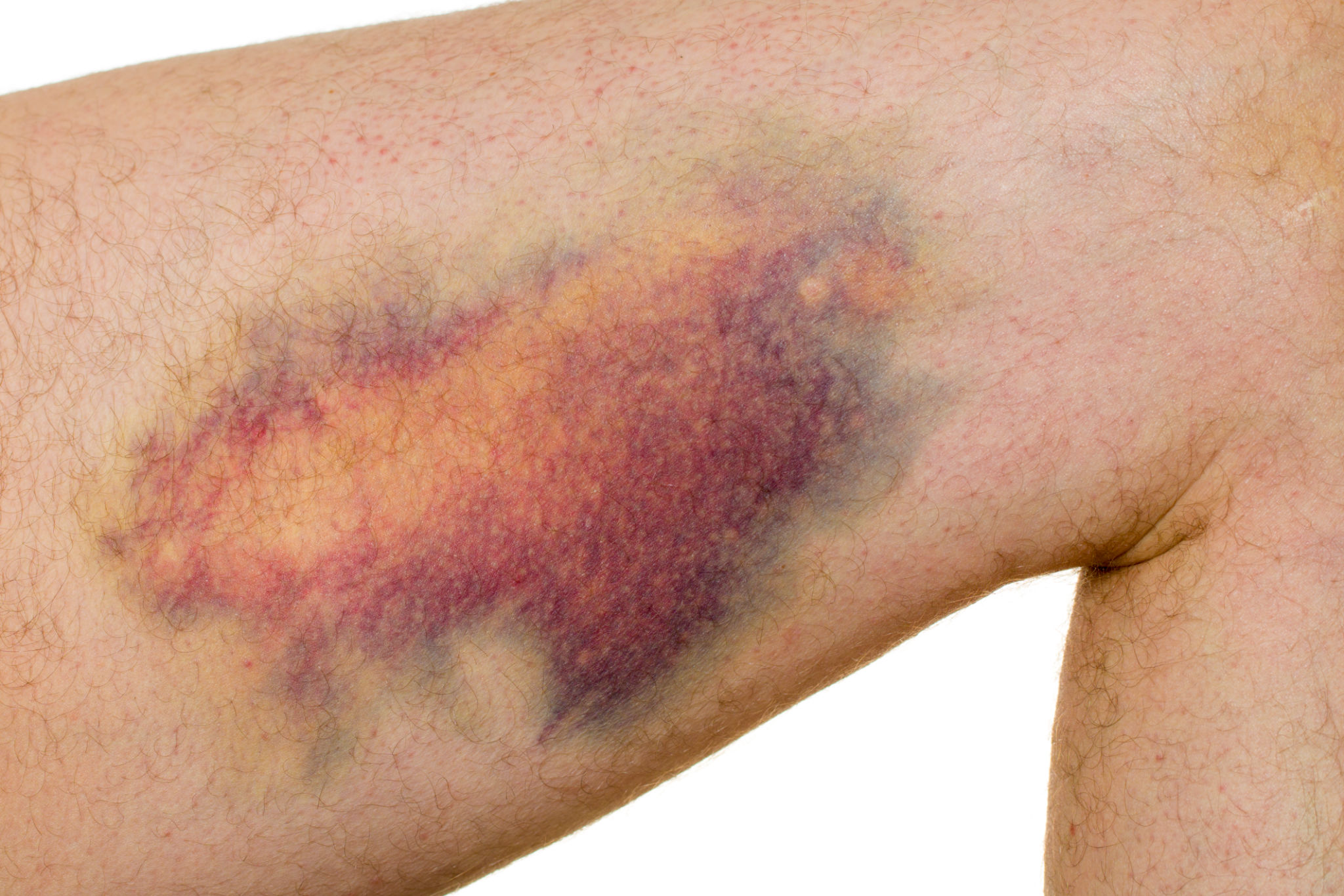
Muscles of our body perform several vital functions such as helping us walk, stand and general mobility. In other scenarios like sports where extreme performance of the muscles is undertaken injuries such as muscle tears are common. Haematoma is a type of swelling in the soft tissues that are caused by bleeding.
Muscle tears and haematomas can cause severe pain, swelling, redness, limited mobility and in some cases difficulty to weightbear and walk.
Initial treatment can include rest, ice packs, limb elevation as appropriate and analgesia. However, when symptoms do not subside or in complex injuries, a systematic assessment from sports injury experts is important to prevent secondary complications like scarring, contracture and chronic pain. Apart from conventional treatment Dr Darshan Angadi uses image guided therapies in some cases to help restore the muscle function and ensure pain free recovery.
5.5 Ligament injuries and sprains

Ligaments are vital structures in our body that connect two bony parts. Hence, they are very important in joint stability and function. Ligaments are prone to injuries and sprains during routine activities or from sports like cricket, football, hockey, rugby etc.
Ligament injuries can result in pain, swelling and restriction in joint movement. In some cases, instability can occur and contribute to weakness and poor balance.
Following a suspected ligament injury, it is often advised to take rest, apply hot/cold compress, elevate the limb as needed and medications in the form of anti-inflammatories. Definitive treatment of ligament injuries requires a good clinical evaluation from a specialist. Subsequently tests and scans may be needed to accurately grade the severity of injury and start precise treatment plan.